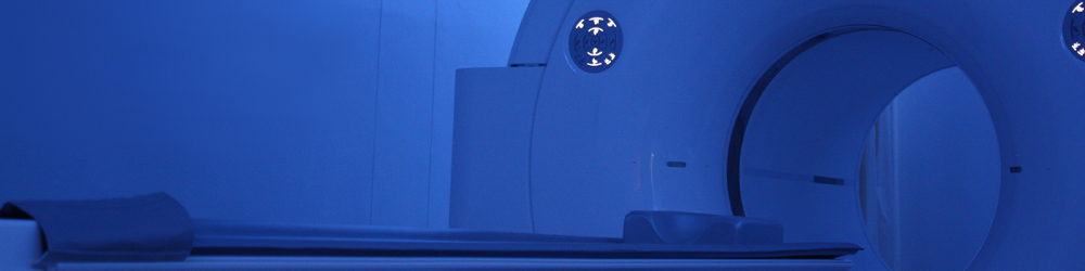
PET (positron emission tomography) is a type of molecular imaging. Molecular imaging allows us to see how the body is functioning at a cellular level and enables measurement of its chemical and biological processes. PET involves using a small amount of injected radioactive substance, known as a radiopharmaceutical, to form a functional image of tissues, normal and abnormal (e.g. cancer or infection) within your body. The radiopharmaceutical is injected intravenously and is taken up by your cells. The PET camera is used to detect the radiation that is emitted from the radiopharmaceutical in your body, this information is used to convert the detected activity into an image.
When it is combined with CT (computed tomography) it is called a PET/CT. We use the CT component to ensure accurate localisation of the uptake seen on the PET. PET/CT is thus a dual imaging modality; this means that we image the cellular/metabolic function (how the cell works) as well as the anatomy (what it looks like) in a single investigation.
The PET/CT scan plays a specific role in the management of your disease and does not replace a CT, MRI or other anatomical imaging. Information gained on these scans complement each other, one scan is not superior to the other. A PET/CT scan may provide information that cannot be obtained from an MRI or vice versa. Your referring doctor will know when a PET/CT scan is the preferred imaging modality.
A big advantage of PET is that it can detect early onset of disease before it is evident on other imaging modalities, i.e. it may detect spread of cancer earlier than on other imaging. Another unique role is using PET to distinguish between dead scar tissue from a treated tumour and active tumour cells, which will have an important influence on your treatment plan. A PET scan may also be used to assess whether your current treatment regime (e.g. chemotherapy or immunotherapy) is effective.